779
Views & Citations10
Likes & Shares
Purpose: To determine the clinical, pathological and biological features of sinonasalpapillomas as they present in two medical centers in Israel.
Materials & methods: Multicenter retrospective cohort study.
Results: Exophytic sinonasalpapillomas were rare. Inverted papillomas, localized exclusively in paranasal sinuses were more frequent than expected. Squamous cell carcinoma developing ex inverted papillomas was found in 7 cases (14.6%), 5 of these patients died of the malignancy. Tumors, including papillomas and squamous cell carcinoma expressed p53 more often than their neighbouring normal mucosa cells. Apoptotic index was higher in benign than in malignant tumors. TGF-β was moderately overexpressed in inverted papillomas.
Conclusions: The higher expression of p53 in tumor tissues is as expected. This is also the case of apoptotic index, which was significantly higher in benign tumors, as compared with carcinoma. This study suggests that TGF-ß1 may play a role in the endophytic pattern of growth of inverted papillomas.
Keywords:Sinonasal Papilloma, Inverted Papilloma, Squamous Cell Carcinoma, TGF-ß1, Apoptotic Index
Abbreviations:TGF-beta: Transforming growth factor-beta;IP: Inverted papilloma; SCC: Squamous cell carcinoma; HPV: Human papilloma virus; TUNEL analysis: TdT-mediated dUTP-FITC nick end labeling; AI: Apoptotic index
INTRODUCTION
Sinonasalpapillomas are benign epithelial tumors composed of respiratory epithelium, often showing squamous metaplasia. Of the three types of sinonasalpapillomas, we will discuss only the two that arise from the lateral nasal walls or from the maxillary sinuses. These are the inverted (IP) and the exophytic papillomas [1].
The endophytic growth of inverted papillomas into the stroma does not disrupt the basement membrane that separate the two. Other unique features of this tumor are a propensity to recur and a tendency to be associated with squamous cell carcinoma (SCC) [1].
The pathogenesis of IP is still obscure. Human papilloma virus (HPV) has been demonstrated in a small number of IP cases only [1]. On the other hand, HPV has been observed, together with p53 overexpression in IP that are synchronous with SCC [2].
The significance of an independent p53 overexpression, as well as that of a high Ki-67 proliferation index, has not been completely clarified to date in regarding IP.
In order to elucidate the endophytic mode of growth of IP, we suggest a possible role for transforming growth factor (TGF)-ß1. This growth factor overexpression is thought to modulate the interaction between epithelium and underlying matrix and may be the cause of the ingrowth of theepithelium in IP [3,4].
MATERIALS & METHODS
Patients with inverted or exophytic papillomas were identified from the archives of the Departments of Pathology of the Soroka Medical Center in Beer-Sheva (Southern Israel) and of the Tel-Aviv Sourasky Medical Center (Central Israel). The diagnosis of the two types of tumor, as well as that of the SCC, was confirmed by two pathologists and a representative section was chosen for immunohistochemistry (IHC) and for the TUNEL assay.
The IHC study was carried out on paraffin-embedded tissues using the avidin-biotin peroxidase complex method with the Vectastain kit of Vector Laboratories (Burlingame, CA, USA), as described [5]. The antibodies used included anti-p53, clone DO-7 (Dako, Glostrup, Denmark) at concentration 1:200; Ki-67/MIB-1 (Dako, Glostrup, Denmark) and TGF-ß1 rabbit, polyclonal antibody (1:200) (Santa Cruz, CA, USA).
The TUNEL assay: apoptosis was studied by the TdT-mediated dUTP-FITC nick end labeling (TUNEL) assay. DNA fragments generated by apoptosis were detected by the ApopTag apoptosis detection kit from Chemicon (Millipore, Billerica, MA, USA) as used previously [5]. We calculated an apoptotic index (AI) as the proportion of apoptotic cells out of a total of 300 benign or else tumor cells.
STATISTICAL ANALYSIS
Descriptive statistics of the study sample were used to summarize participant characteristics. Mann-Whitney U-test was used for comparison of two non-parametric means, different proportions were analyzed in cross tables using Fisher's exact test. For evaluation of associations between continuous variables, bi-variate Spearman’s correlations were conducted.
All tests of significance were two-tailed with level of significance
RESULTS
Among 51 cases with sinonasalpapillomas, 41 (80.4%) were diagnosed as inverted papilloma. In 3 cases, both patterns (inverted and exophytic papillomas) were found. In 7 cases, inverted papilloma was associated with SCC.Exophytic papilloma-only was diagnosed in a single case.
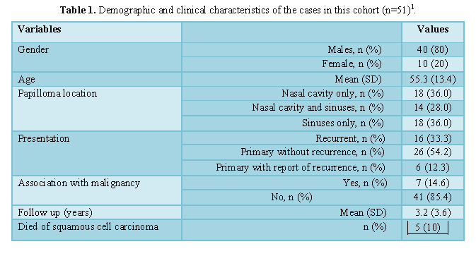
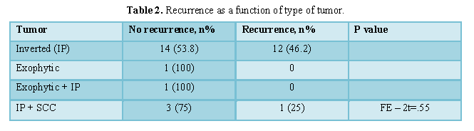
The age at diagnosis ranged between 22 and 86 years (mean 55.3+/-13.4 years). Eighty percent of the patients were men. The mean time of follow-up was 3.2 years. In 36% of cases, the papilloma involved the nasal cavity only. Papillomas involving the paranasal sinuses exclusively were found in 36% of the cases. In 28% of cases, both nasal cavity and sinuses were involved. Twenty-two cases (45.8%), mainly IP, were associated with recurrence of the lesion. Seven (14.6%) of the inverted papilloma were associated with SCC. Of these, five patients died of the SCC. The demographic and clinical features are summarized in (Tables 1 and 2).
The surgical procedures for the intranasal lesions included polypectomy, functional endoscopic sinus surgery and ethmoidectomy. Mid-facial de-gloving, medial maxillectomy and the Caldwell-Luc approach were carried out for extra nasal lesions.
Forty-one papillomas were available for IHC analysis. Thirty-two of these (78%) were inverted, one exophytic and two mixed. In five cases (12.2%), inverted papilloma was found within SCC. The follow-up of this group ranged from 0.00 to 14 years. Thirteen samples of recurrent papilloma were analyzed by IHC.
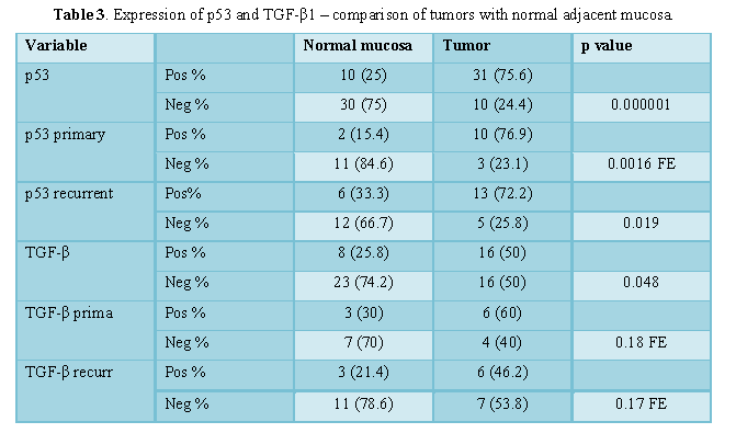
Comparison of the marker’s expression in the primary tumor with that of the adjacent normal epithelium and the recurrent tumor is shown in (Table 3). Tumors exhibited a significantly higher expression of p53 and TGF-β1 in comparison with normal adjacent epithelium.
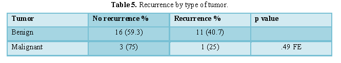
Benign tumors were associated with a significantly higher AI when compared with malignant tumors (Tables 4 and 5).
When considering the sample of 41 inverted papillomas, we found statistically significant correlations between Ki-67 as expressed in adjacent normal tissues with Ki-67 expressed in the tumors (p<.001). A significant correlation was noted between Ki-67 as expressed in IP with the AI of IP (p= .041). In addition, all the above was significant for the IP with recurrence. We were unable to demonstrate significant differences between the two medical centers with regard to features of sinonasalpapillomas (data not shown).
DISCUSSION
The population sampled from our two medical centers stands out for the limited number of exophytic nasal papilloma’s. When this type of papilloma occurred, it tended to be admixed with inverted papillomas. This has not been the experience of most previous studies [6-8].
Papillomas, mainly of inverted type and involving exclusively the paranasal sinuses represent 36% of our cohort - a more frequent occurrence than previously described [9]. The rate of recurrence (45.8%) is also higher than expected. It predominantly concerned the IP. The recurrence of benign tumors, notably of inverted papilloma is superior to that of SCC ex inverted papilloma, though not to a significant degree [10,11]. Mortality from SCC associated with IP (10%) is probably higher than previously reported [12,13].
We investigated the expression of p53 in the various tissues. Tumor tissues showed a significantly higher expression ofp53. The HPV status of these tumors is not available, as it may be relevant for IP associated with SCC only [2,14]. Moreover, normal mucosa was significantly more negative in non-recurrent tumors, while p53 was more positive in recurrenttumors, but not to a significant degree. Our findings sustain the view that for its immunohistochemical expression, the p53 antigenic product should be related with either a mutated or with a deleted gene (functionally, under the influence of a virus or Mdm-2, or otherwise).
Apoptotic index was found to be significantly higher in benign tumors than in SCC and this result is expected [15,16].
Ki-67 index showed a correlation between the normal mucosa and the IP. This may be an indirect evidence of a field effect. A significant correlation was found between Ki-67 and AI within tumors. These findings need clarifying as a strong correlation between a high proliferation fraction and activated apoptosis is typical of very high-grade tumors [15,16], like Burkitt lymphoma.
TGF-β1 was chosen in our study for the putative relevance it may have in the reverse mode of growth in IP. With this regard, a possible role for TGF-β1 in the interaction between the epithelium and the matrix may be expected [3,4]. In fact, TGF-β1 was significantly more negative in normal adjacent mucosa, while it was relatively overexpressed (p=0.048) in tumor tissue, mainly in the IP. This pathogenetic avenue should be explored further, also in conjunction with inverted urothelial papilloma’s and with the inverted growth of urothelial carcinoma.
CONCLUSIONS
The morphological, clinical and laboratory characteristics of nasal papilloma’s from two distinct areas of Israel are described. A restricted number of exophytic nasal papilloma’s were observed. The exclusive localization of IP in paranasal sinuses is more frequent than expected. Tumor tissues showed a higher expression of p53 than the neighboring mucosa. Benign tumors show a higher apoptotic index than SCC. TGF-β1 overexpression is proposed to represent the mechanism by which the endophytic growth pattern of inverted papilloma develops.
ACKNOWLEDGEMENTS
This study was funded in part by the Israel Ministry of Health. We are grateful to Eugenia Mejirovski for her excellent technical work.
CONFLICT OF INTERESTS
None. The institutional "Helsinki" Committee approval: SOR-0100-12.
1. Krishnappa PK (2009) Occult papillary carcinoma thyroid presenting as lateral cystic neck mass-A case report & review of literature. Int J Pathology 10.
2. Buchwald C, Bradley PJ (2007) Risks of malignancy in inverted papilloma of the nose and paranasal sinuses. CurrOpinOtolaryngol Head Neck Surg 15: 95-98.
3. Buchwald C, Lindeberg H, Pedersen BL, Franzmann MB (2001) Human papilloma virus and p53 expression in carcinomas associated with sinonasalpapillomas: A Danish epidemiological study 1980-1998. Laryngoscope 111: 1104-1110.
4. Silberstein GB, Strickland P, Coleman S, Daniel CW (1990) Epithelium-dependent extracellular matrix synthesis in transforming growth factor-β1-growth inhibited mouse mammary gland. J Cell Biol 110: 2209-2219.
5. Cardillo MR, Petrangeli E, Salvatori L, Ravenna L, Di Silverio F (2000) Transforming growth factor-β1 and androgen receptors in prostate neoplasia. Anal Quant CytolHistol 22: 403-410.
6. Benharroch D, Levy A, Prinsloo I, Ariad S, Rabinovitch D, et al. (1999) Apoptotic index as a prognostic factor in Hodgkin’s disease. Leuk Lymph 33: 351-359.
7. Franzmann MB, Buchwald C, Jacobsen GK, Lindeberg H (1998) Expression of p53 in normal nasal mucosa and in sinonasal papilloma with and without association with carcinoma and the relation to human papilloma virus (HPV). Cancer Lett 128: 161-164.
8. Buchwald C, Franzmann MB, Tos M (1995) Sinonasal papilloma: A report of 82 cases in Copenhagen County, including longitudinal epidemiological and clinical study. Laryngoscope 105: 72-79.
9. Norris HJ (1962) Papillary lesions of the nasal cavity and paranasal sinuses. I Exophytic (squamous) papilloma. A study of 28 cases. Laryngoscope 72: 1784-1797.
10. Eggers G, Muhling J, Hassfeld S (2007) Inverted papilloma of the paranasal sinuses. J Craniomaxillofac Surg 35: 21-29.
11. Kim YM, Kim HS, Park JY, Koo BS, Park YH, et al. (2008) External versus endoscopic approach for inverted papilloma of the sinonasal cavities: A retrospective study of 136 cases. Acta Otolaryngol 128: 909-914.
12. Sham CL (2009) Treatment results of sinonasal papilloma: An 18-year study. Am J RhinolAllerg 23: 203-211.
13. Mendelhall WH, Hinerman RW, Malyapa RS, Werning JW, Amdur RJ, et al. (2007) Inverted papilloma of the nasal cavity and paranasal sinuses. Am J Clin Oncol 30: 560-563.
14. Tanvetyanon T, Qin D, Padhya T, Kapoor R, McCaffrey J, et al. (2009) Survival outcomes of squamous cell carcinoma arising from sinonasal inverted papilloma: Report of 6 cases with systematic review and pooled analysis. Am J Otolaryngol 30: 38-43.
15. Altavilla G, Staffier A, Busatto G, Canesso A, Giacomelli L, et al. (2008) Expression of p53, p16(INK4A), pRb, p21(WAF1/CIP1), p27(KIp1), cyclin D1, Ki-67 and HPV DNA in sinonasal endophytic Schneiderian (inverted) papilloma. Acta Otolaryngol 31: 1-8.
16. Fan GK, Imanaka M, Yang B, Takenaka H (2006) Characteristics of nasal inverted papilloma and its malignant transformation: A study of cell proliferation and programmed cell death. Am J Rhinol 20: 360-363.
17. Katori H, Nozawa A, Tsukuda M (2007) Cell proliferation, apoptosis and apoptosis inhibition in malignant transformation of sinonasal inverted papilloma. Acta Otolaryngol 127: 540-546.
-
 Table 1
 -
Table 2
-
Table 3
-
Table 4
-
Table 5

QUICK LINKS
- SUBMIT MANUSCRIPT
- RECOMMEND THE JOURNAL
-
SUBSCRIBE FOR ALERTS
RELATED JOURNALS
- International Journal of Medical and Clinical Imaging (ISSN:2573-1084)
- Journal of Pathology and Toxicology Research
- Journal of Psychiatry and Psychology Research (ISSN:2640-6136)
- Archive of Obstetrics Gynecology and Reproductive Medicine (ISSN:2640-2297)
- BioMed Research Journal (ISSN:2578-8892)
- International Journal of Internal Medicine and Geriatrics (ISSN: 2689-7687)
- Advance Research on Endocrinology and Metabolism (ISSN: 2689-8209)


